Researchers Unveil Groundbreaking Technique in X-Ray Imaging
Technology Could Lead to Improvements in Medical Diagnostics, Materials and Industrial Imaging, Transportation Security
Researchers at the University of Houston unveiled a groundbreaking advancement in X-ray imaging technology that could provide significant improvements in medical diagnostics, materials and industrial imaging, transportation security and other applications.

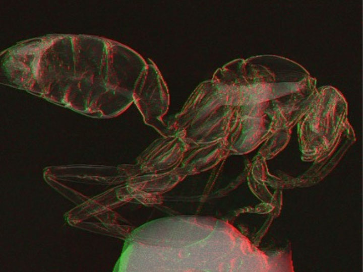
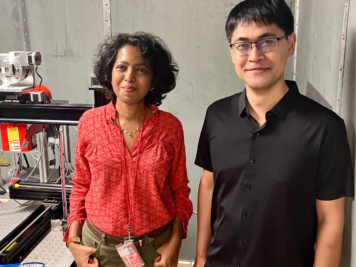
In a paper featured on the cover of Optica, one of the world’s leading journals in theoretical and applied optics and photonics, Mini Das, Moores Professor of Physics at UH’s College of Natural Sciences and Mathematics and Cullen College of Engineering, and Jingcheng Yuan, a physics graduate student, introduce a novel light transport model for a single-mask phase imaging system that enhances non-destructive deep imaging for visibility of light-element materials, including soft tissues such as cancers and background tissues like plastics and explosives.
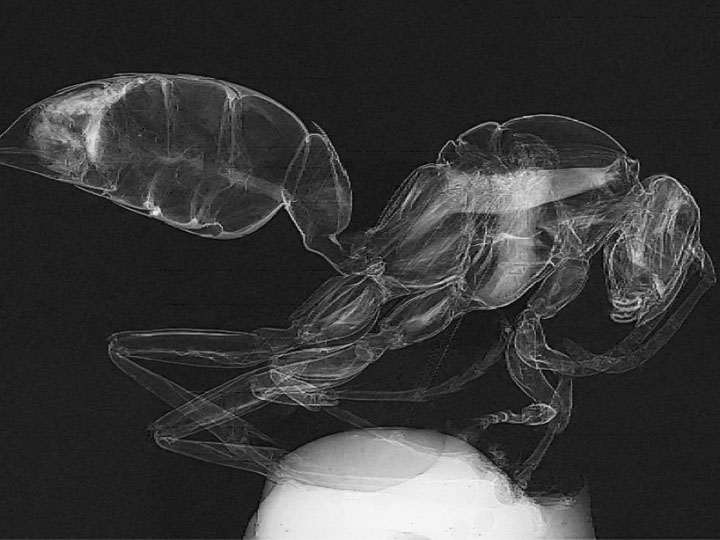
“Older X-ray technology relies on X-ray absorption to produce an image,” Das said. “But this method struggles with materials of similar density, leading to low contrast and difficulty distinguishing between different materials, which is a challenge across medical imaging, explosive detection and other fields.”
Enhanced Contrast for Soft Tissues

X-ray phase contrast imaging, or PCI, has gained considerable attention in recent years for its potential to provide enhanced contrast for soft tissues by utilizing relative phase changes as the X-rays go through an object. Among many techniques available, the single-mask differential stands out for its simplicity and effectiveness to translate to practical applications and in yielding higher contrast images compared to other methods. And it does so in a much more simple and efficient way with single-shot, low-dose imaging.
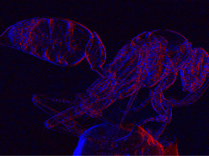
“Our new light transport model enables the understanding of contrast formation and how multiple contrast features mingle in acquired data,” Das said. “As a result, it allows the retrieval of images with two distinct types of contrast mechanisms from a single exposure, which is a significant advancement over traditional methods.”
The design uses an X-ray mask with periodic slits, creating a compact setup that enhances edge contrast.


“This mask aligns with detector pixels, allowing us to capture differential phase information which shows variations between materials more clearly. The main advantage of this is that it simplifies the setup and reduces the need for high-resolution detectors or complex, multi-shot processes.”
Goal: Integrate Technology into Portable Systems for Testing
Das’s team has already tested its model via rigorous simulations and on their in-house developed laboratory benchtop X-ray imaging system. The next goal, she says, is to integrate the technology into portable systems and retrofit existing imaging setups to test it in real-world environments, such as hospitals, industrial-ray imaging and airports.
“Our research opens up new possibilities for X-ray imaging by providing a simple, effective and low-cost method for enhancing image contrast which is a critical need for non-destructive deep imaging,” Das says. “It makes phase contrast imaging more accessible and practical, leading to better diagnostics and improved security screening. It is a versatile solution for a wide range of imaging challenges. We are in the process of testing the feasibility for a number of applications.”
Das’s research is funded through multiple agencies including the National Science Foundation, Congressionally Directed Medical Research Programs, and National Institutes of Health. The latest funding from the National Institute of Biomedical Imaging and Bioengineering aims to develop low-dose Micro-CT that utilizes multiple novel contrast mechanisms, thereby reducing radiation dose and imaging time which continues to be a significant issue.
Bryan Luhn, University Media Relations

