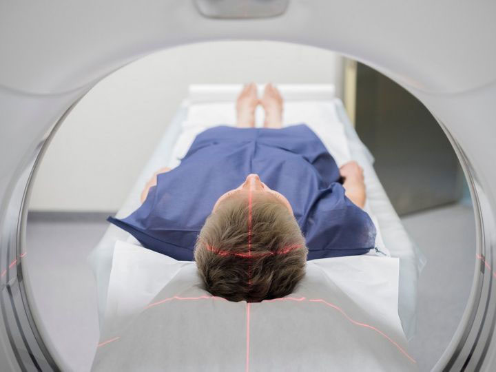
Next-Gen Micro-CT Scan Can Lower Radiation, Offer Better Pictures
Research Could ‘Transform the Landscape’ of Micro-CT Imaging
With a five-year, $3.2 million grant from the National Institute of Biomedical Imaging and Bioengineering, Mini Das, associate professor of physics at the University of Houston, will help usher in the next generation of micro computed tomography (CT) imaging.

The project’s goal is to lower radiation dose in X-ray micro-CT imaging while improving the resolution and enhancing the contrast of three-dimensional pictures of small specimens, like tumors or biomaterials.
“This has the potential to transform the landscape of micro-CT imaging,” said Das, who recently developed the theory, instrumentation and algorithms for spectral phase-contrast imaging (PCI) to enable the use of much lower doses of radiation while delivering higher levels of image detail.
Challenges of Current Micro-CT Scanning
Das’s work addresses the challenge of current in-vivo micro-CT scanning – long imaging times, harmful, yet required, high radiation dose levels needed to follow the same subject over time, and poor image contrast.
“Current X-ray and CT systems have inherent contrast limitations, and dense tissue and cancer can often look similar. Even if you increase the radiation dose, there is a limit to what you can see. In addition, image noise becomes significant when increasing resolution to see fine details, often desirable when scanning small objects,” said Das, who is on the faculty of the UH College of Natural Sciences & Mathematics.
Higher Contrast Between Tissue Types, Improved Images
PCI detects how X-rays bend, or refract, particularly at the interface between tissues, providing a higher amount of contrast between different types of tissue. It also measures absorption and phase changes from X-ray transmission through the body. The resulting phase-enhanced contrast depicts high fine-structure visibility adding new image features. The developed methods can also translate to large-scale CT systems.
Detecting the bending of X-rays is challenging because the bend is small, and they are both bending and being absorbed in tissues at the same time, complicating interpretation.
“X-rays, like visible light, exhibit what is called dual nature - they behave both as particles, called photons or packets of light, and waves. Phase imaging methods capture information relevant to wave nature of X-rays unlike conventional imaging systems found in clinics today,” said Das.
Goal: Higher Levels of Image Detail
Das is using a new multi-energy, or spectral, detector which can see the energy of every light particle and will develop a unique spectral micro-CT system with both PCI and non-PCI capabilities.
“We are the first to show how to use photon-counting detectors in a phase-contrast imaging setting while extracting the absorption and phase effects with quantitative accuracy. This accurate phase retrieval, or recovery, is so important if you want to discriminate between cancers and normal tissues,” she said.
Multiple patents and publications from her group have shown significant preliminary data and feasibility for the new methods.
Both physics and engineering students will work together on the project, which involves international collaboration. Das also has a dual appointment in physics and biomedical engineering allowing an interdisciplinary and collaborative approach to her work.
Laurie Fickman, University Media Relations

